What is an Echocardiogram?
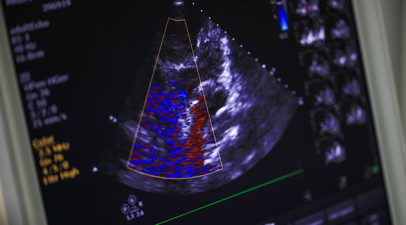
Echocardiography is a test that uses sound waves to produce live images of your heart. The image is an echocardiogram. This test allows your doctor to monitor how your heart and its valves are functioning. The images can help them spot:
- Blood clots in the heart.
- Fluid in the sac around the heart.
- Problems with the aorta, which is the main artery connected to the heart.
An echocardiogram is key in determining the health of the heart muscle, especially after a heart attack. It can also reveal heart defects in unborn babies.
Taking an echocardiogram is painless. There are only risks in very rare cases with certain types of echocardiograms.
Your doctor may order an echocardiogram for several reasons. For example, they may have discovered an abnormality from other testing or while listening to your heartbeat through a stethoscope. If you have an irregular heartbeat, your doctor may want to inspect the heart valves or chambers or check your heart’s ability to pump. They may also order one if you’re showing signs of heart problems, such as chest pain or shortness of breath.
- Bladder
- Brain (in infants)
- Eyes
- Gallbladder
- Kidneys
- Liver
- Ovaries
- Pancreas
- Spleen
- Thyroid
- Testicles
- Uterus
- Blood vessels
There are several different types of echocardiograms.
Transthoracic Echocardiography
This is the most common type of echocardiography. It’s painless and noninvasive.
A device called a transducer will be placed on your chest over your heart. The transducersends ultrasound waves through your chest toward your heart. A computer interprets the sound waves as they bounce back to the transducer. This produces the live images that are shown on a monitor.
Transesophageal echocardiography
If a transthoracic echocardiogram doesn’t produce definitive images, your doctor may recommend a transesophageal echocardiogram. In this procedure, the doctor guides a much smaller transducer down your throat through a thin, flexible tube in your mouth. They will numb your throat to make this procedure easier.
This tube is guided through your esophagus, the tube that connects your throat to your stomach. With the transducer behind your heart, your doctor can get a better view of any problems.
Stress Echocardiogram
A stress echocardiogram uses traditional transthoracic echocardiography. However, the procedure is done after you’ve exercised or taken medication to make your heart beat faster. This allows your doctor to test how your heart performs under stress.
Three-dimensional Echocardiography
A three-dimensional (3-D) echocardiogram uses either transesophageal or transthoracic echocardiography to create a 3-D image of your heart. This involves multiple images from different angles. It’s used prior to heart valve surgery. It’s also used to diagnose heart problems in children.
Fetal Echocardiography
Fetal echocardiography is used on expectant mothers sometime during weeks 18 to 22 of pregnancy. The transducer is placed over the woman’s belly to check for heart problems in the fetus. The test is considered safe for an unborn child because it doesn’t use radiation, unlike an X-ray.
Echocardiograms are considered very safe. Unlike other imaging techniques, such as X-rays, echocardiograms don’t use radiation.
A transthoracic echocardiogram carries no risk. There’s a chance for slight discomfort when the electrodes are removed from your skin. This may feel similar to pulling off a Band-Aid.
There’s a rare chance the tube used in a transesophageal echocardiogram may scrape the side of your esophagus and cause irritation. The most common side effect is a sore throat. You may also feel a bit funny due to the sedative used in the procedure.
The medication or exercise used to get your heart rate up in a stress echocardiogram could temporarily cause an irregular heartbeat. The risk of a serious reaction is reduced because the procedure is supervised.
A transthoracic echocardiogram requires no special preparation.
However, if you undergo a transesophageal echocardiogram, your doctor may instruct you not to eat anything for a few hours before the test. This is to prevent you from vomiting during the test. You may also not be able to drive for a few hours afterward due to the sedatives.
If your doctor has ordered a stress echocardiogram, wear clothes and shoes that are comfortable to exercise in.
Your doctor will review your results after the test. The results may reveal abnormalities such as:
- Damage to the heart muscle
- Heart defects
- Heart size
- Pumping strength
- Valve problems
If your doctor is concerned about your results, they may refer you to a cardiologist. This is a doctor who specializes in the heart. Your doctor may order more tests or physical exams before diagnosing you.
If you’re diagnosed with a heart condition, your doctor will work with you to develop a treatment plan that works best for you.